Search
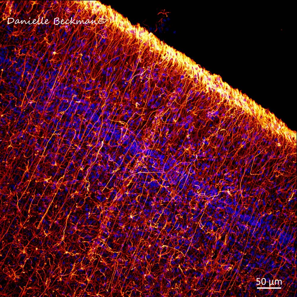
- How do neurons know their final position in the brain? They use a combination of chemical signals & physical guidance by radial glia 🟡. My favorite cell type in the brain is also temporary! After cortical development, most will transform into astrocytes. Nuclei of cells in🔵 #SciArt #Microscopy
- 💡A must-read: how can we ensure microscopy tools are used, not just shipped, in underserved regions? Brilliant insights in this new Nature Methods Comment. 🔗https://www.nature.com/articles/s41592-025-02690-7 #Microscopy #ScienceEquity
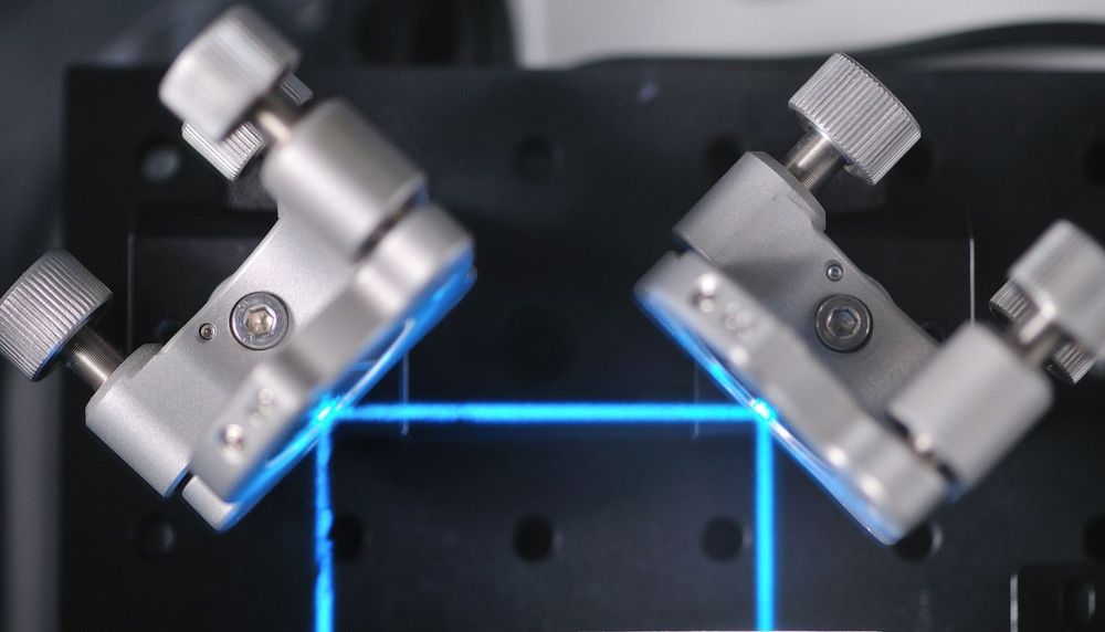
- ONI’s #AploScope #SuperResolution #microscope has been engineered for reliability and maximal performance. Its powerful imaging capabilities and integrated software help ensure reproducibility across experiments and users. Discover the power of Aplo! oni.bio/aploscope #microscopy #SMLM #dSTORM
- Deadline extended to 19 May! 🧪 Learn about light sheet #microscopy, sample preparation, data processing and #ImageAnalysis. Apply now for EMBO Practical Course on Light sheet microscopy in Dresden, Germany, 11–22 August meetings.embo.org/event/25-lsm #EMBOlsm #imaging #EMBOevents
- Cleary, Qiu, Looney et al. report an intravital #microscopy approach for imaging lymphatic vessels in the lungs. This technique can also reveal cellular dynamics in other understudied tissue structures rupress.org/jem/article/... 📘 In JEM #Immunology Collection: rupress.org/jem/collecti... #AAI2025
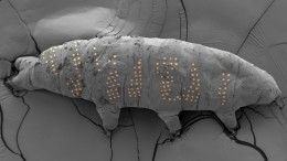
- A tardigrade just got the world's tiniest tattoo. This, and so much more, in Nature's roundup of the best #science images from April. 🧪 And yes, you should take 20 seconds to play the aurora video. 😍 www.nature.com/immersive/d4... #SciencePhotography #Microscopy
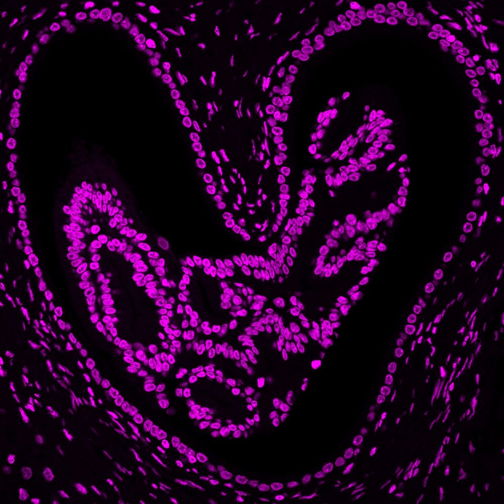
- A love heart for you all, this sunny #FluorescenceFriday. Pass it on to show some love to your friends who love #Microscopy ❤️🔬
- A new mushroom microscopy video just came out - this goes into detail on how to prepare microscope slides and find various taxonomically significant features such as spores, cheilocystidia, pleurocystidia, pileipellis and more. youtube.com/watch?v=vMY2... #fungifriends #fungi #microscopy
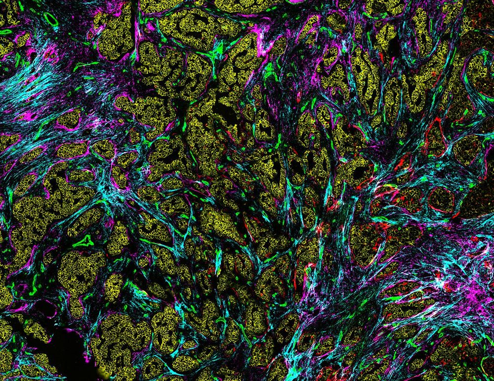
- This image, from a clinical study called TRACERx, shows the molecules organised outside cells that act as a scaffold in tissues. These molecules (red, green, cyan and magenta) are highly modified in cancer. The tumour cells are depicted in yellow 🧪 #microscopy